Skip to content
Skip to content
  • News
  • Events
  • eBooks
  • Blog
  • Careers
  • Contact
  • News
  • Events
  • eBooks
  • Blog
  • Careers
  • Contact
KNOWLEDGEBASE
  • About Us
  • Products

    Fluorescence Spectrometers

    • FLS1000 Photoluminescence Spectrometer
    • FS5 Spectrofluorometer
    • LifeSpec II Lifetime Spectrometer
    • Mini-tau Lifetime Spectrometer

    Raman Microscopes

    • RM5 Raman Microscope
    • RMS1000 Multimodal Confocal Microscope

    Transient Absorption

    • LP980 Transient Absorption Spectrometer

    FTIR Spectrometers

    • IR5 FTIR Spectrometer

    Lasers and LEDs

    • Pulsed Lasers
    • Gas Lasers
    • Customisation Options
    View All Products
  • Techniques
  • Applications
KNOWLEDGEBASE
Edit Content
  • About Us
  • Products
  • Techniques
  • Applications
  • Knowledgebase
  • eBooks
  • News
  • Events
  • Blog
  • Careers
  • Contact Us

RESOURCES

3D Raman Mapping of a Transdermal Patch

  • September 9, 2021

KEY POINTS

  • Transdermal patches deliver drugs through the skin into the bloodstream, providing controlled medication release.
  • 3D Raman mapping can be used to analyse the composition of transdermal patches, revealing the active pharmaceutical ingredient (API) and polymer layers.
  • This technique is useful for quality control in pharmaceutical laboratories, ensuring correct production and monitoring the stability of adhesive layers.
  • The analysed nicotine patch was found to contain six layers, including poly(ethylene terephthalate) (PET) and polyethylene (PE), with nicotine embedded in a polymer reservoir.
Edit Content

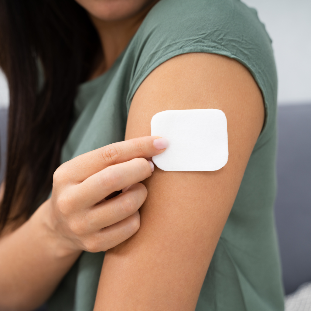
A transdermal patch is a method of drug delivery through the skin into the bloodstream. The patch contains a drug which absorbs into your skin when attached to you and it releases a consistent and controlled amount of medication into the body over long periods time. This method of delivery is preferred in cases where the drug can have negative side effects on the gastrointestinal tract, for prolonged drug release, or an option for people with a fear of needles looking to avoid medication delivered via injection.

layers in a transdermal nicotine patch

Figure 1: Typical transdermal patch and its layer composition.

One of the most common uses for transdermal patches is to help combat nicotine addiction. Nicotine transdermal patches were first developed as a method to aid smokers quitting in the 1980s after several studies proved the absorption of nicotine into the body reduced cravings. Nicotine replacement therapy (NRT) releases small amounts of nicotine throughout the day, without the additional harmful components of cigarettes, such as tar. A recent review of data from the public and from clinical trials revealed using an NRT can increase the chance of successfully quitting by up to 60%.1

In this application note we study a nicotine patch to reveal the active pharmaceutical ingredient (API) as well as the polymer layers encapsulating it via confocal Raman microscopy. Using an RMS1000 Raman Microscope we analysed a nicotine patch in the X, Y, and Z dimensions. This can provide manufacturers and researchers with important information on the quality of their product, validating the amount and distribution of API within the patch, as well as identifying any contaminants without causing damage to the sample.


Materials and Methods

An RMS1000 Raman Microscope was used for the 3D measurement of the transdermal nicotine patch. The nicotine patch was a commercially available NRT purchased from a pharmacy. The RMS1000 can take 3D Raman maps thanks to its confocal pinhole. The pinhole acts as a spatial filter in the axial (Z) direction by blocking the Raman scatter from above and below the focal plane and is essential for 3D mapping. Without the pinhole, there is no axial spatial filtering and Raman scatter from the entire axial volume of the sample would be collected with no axial resolution.

Figure 2: RMS1000 Raman Microscope.

The RMS1000 has several confocal pinhole positions available, from 25 µm to 2 mm, the smaller the confocal pinhole the better the resolution in X, Y, and Z. However, as you decrease the pinhole you will decrease the Raman intensity, therefore, by offering many pinhole positions the RMS1000 allows the user to find the perfect balance of spatial resolution and Raman intensity for their sample.

The 3D Raman map of the patch was taken with a 785 nm laser at 100% power and the pinhole set to 300 µm. A slit width of 70 µm was used along with a 600 gr/mm grating and a back illuminated CCD.


Results

The transdermal patch analysed contained nicotine as they active ingredient surrounded by polymers. By carrying out a Raman 3D map we can visualise the different material layers present in patch, shown below in Figure 3 as a 3D render. Spectra were collected at 10 µm steps in the X and Y axis, and 1 µm steps in the Z axis.

3D Raman map of a nicotine transdermal patch

Figure 3: 3D Raman map of a nicotine transdermal patch.


The 3D Raman map, Figures 3 & 4, reveals the patch contains 6 layers, the spectrum of each layer can be extracted and analysed for identification. The outer layers of the patch, layers 1 and 6, shown in red, can be identified as poly (ethylene terephthalate) (PET) using the KnowItAll Raman Database. The 3D map is coloured based on peaks specific to the material identified, for example for the layers of PET the peak at 1293 cm-1 was selected. Layer 2 can be identified as PET/polyisobutylene, labelled in pink, based on the 1233 cm-1 peak and layers 3 and 5 which surround the API were determined to be polyethylene (PE), highlighted in blue from its peak at 1442 cm-1. Finally, in the middle of the polymers is a thick layer of the API, nicotine, embedded in polymer reservoir (ethylene). This is shown in green and is attributed to peaks at 1045 cm-1 and 1030 cm-1. Layer 1 is the release liner of the patch; this is removed before application and is responsible for protecting the medication and adhesive. Layer 2 is the adhesive part of the nicotine patch, layers 3 and 5 are microporous PE which control the release of the API in layer 4. Layer 6 is the backing which provides protection to the medication whist the patch is being worn.

Raman spectra from the layers of a transdermal patch using an RMS1000

Figure 4: Left- Raman spectra from each component. Right – XZ cross section of the sample.


Conclusion

This application note demonstrated how the RMS1000 Raman Microscope can be used for 3D mapping of a nicotine transdermal patch. The API can be visualised, in this case nicotine, as can the surrounding polymers responsible for protecting and ensuring the release of the API. Thanks to the fingerprint like chemical identification offered by Raman spectroscopy the different polymers can be differentiated, and their thickness assessed. In this way confocal Raman Microscopy is a useful technique for pharmaceutical laboratories to quality control their products ensuring they have been produced correctly without contaminants and help monitor the stability of adhesive layers.
 

References

  1. J. Hartmann-Boyce et al., Nicotine replacement therapy versus control for smoking cessation, Cochrane Database Syst. Rev.5, CD000146, (2018)

RELATED PRODUCTS

RMS1000

Multimodal Confocal Microscope

VIEW

RM5

Raman Microscope

VIEW

Contact our expert team today to find out more about how our products can improve your research

Contact Us
PrevPrevious
NextNext
Previous Energy Transfer in a Liquid Scintillator Investigated using Time-Resolved X-ray Excited Luminescence Spectroscopy Next Choosing Laser and LED Sources for Time-Resolved Photoluminescence Experiments

RESOURCES

Tags:
  • Application Notes
  • Biomedical
  • Pharmaceuticals
  • RM5
  • RMS1000
  • Raman
Download PDF
Suggested Reading:

Identification of Microplastics Using Raman Spectroscopy

View more »
Share:

Keep up to date with the latest from Edinburgh Instruments

Join our mailing list and keep up with our latest videos, app notes and more!

LOCATION:
  • Edinburgh Instruments Ltd.
    2 Bain Square, Kirkton Campus, Livingston, EH54 7DQ.
  • sales@edinst.com
  • +44 1506 425 300
ABOUT:
  • About Us
  • Techniques
  • Applications
  • Knowledgebase
  • About Us
  • Techniques
  • Applications
  • Knowledgebase
PRODUCTS:
  • Fluorescence Spectrometers
  • Raman Microscopes
  • UV-Vis Spectrophotometers
  • Transient Absorption
  • FTIR Spectrometers
  • Lasers and LEDs
  • Customisation Options
  • Software
  • Upgrades
  • All Products
  • Fluorescence Spectrometers
  • Raman Microscopes
  • UV-Vis Spectrophotometers
  • Transient Absorption
  • FTIR Spectrometers
  • Lasers and LEDs
  • Customisation Options
  • Software
  • Upgrades
  • All Products
LEGALS:
  • News
  • Events
  • Blog
  • Careers
  • Contact Us
  • Terms and Conditions
  • Privacy Policy
  • News
  • Events
  • Blog
  • Careers
  • Contact Us
  • Terms and Conditions
  • Privacy Policy
SOCIALS:
Youtube Linkedin X-twitter Facebook
©2024 Edinburgh Instruments. Registered in England and Wales No: 962331. VAT No: GB 271 7379 37
Manage Consent
To provide the best experiences, we use technologies like cookies to store and/or access device information. Consenting to these technologies will allow us to process data such as browsing behaviour or unique IDs on this site. Not consenting or withdrawing consent, may adversely affect certain features and functions.
Functional Always active
The technical storage or access is strictly necessary for the legitimate purpose of enabling the use of a specific service explicitly requested by the subscriber or user, or for the sole purpose of carrying out the transmission of a communication over an electronic communications network.
Preferences
The technical storage or access is necessary for the legitimate purpose of storing preferences that are not requested by the subscriber or user.
Statistics
The technical storage or access that is used exclusively for statistical purposes. The technical storage or access that is used exclusively for anonymous statistical purposes. Without a subpoena, voluntary compliance on the part of your Internet Service Provider, or additional records from a third party, information stored or retrieved for this purpose alone cannot usually be used to identify you.
Marketing
The technical storage or access is required to create user profiles to send advertising, or to track the user on a website or across several websites for similar marketing purposes.
Manage options Manage services Manage {vendor_count} vendors Read more about these purposes
View preferences
{title} {title} {title}