Menu

3D imaging of geological samples is an emerging and exciting application of Raman microscopy. By combining the molecular identification capabilities of Raman spectroscopy with the high spatial resolution of confocal microscopy, detailed information about the distribution of different minerals and organic matter within rocks can be gained. The 3D imaging capability of Raman microscopy is particularly useful for studying:
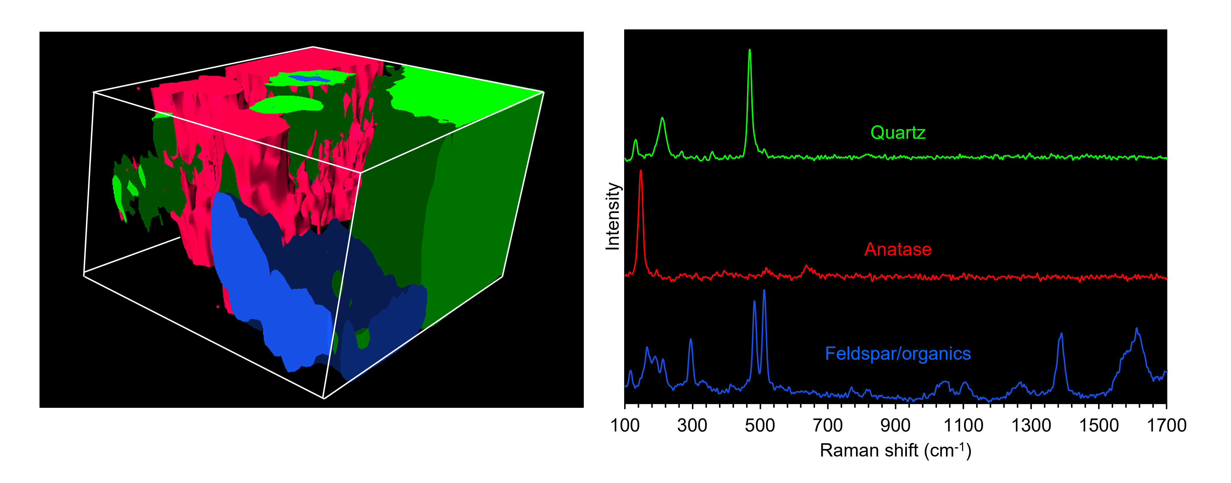
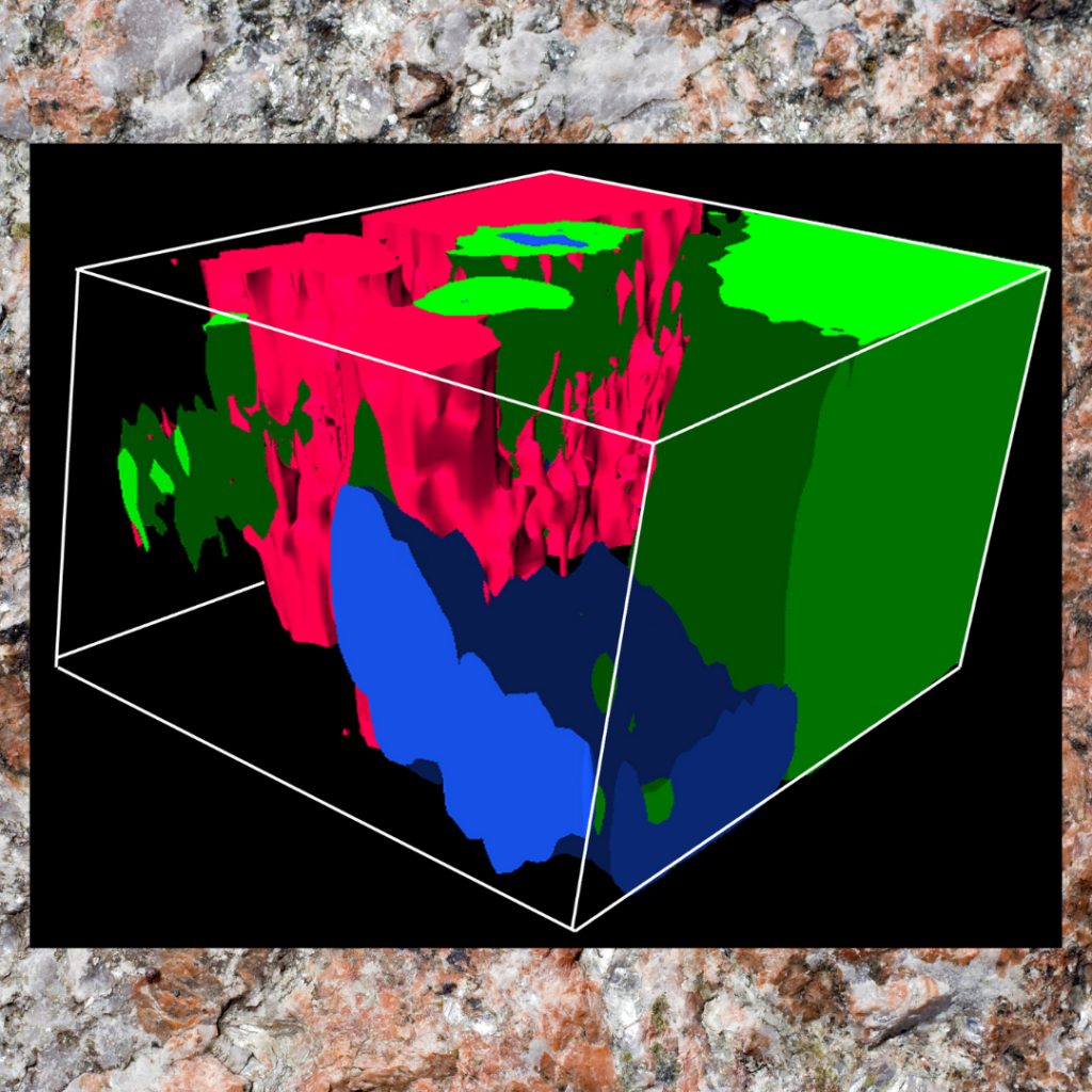
We performed 3D Raman imaging on a crack in a piece of pegmatite rock using the Edinburgh Instruments RM5 Confocal Raman Microscope and identified several components. The result is shown in the image above. We identified quartz and anatase at the top of the crack through their unique Raman signatures. At the bottom of the crack, we found a mixture of feldspar and organics. By combining 3D imaging with detailed chemical information, the RM5 can unravel the complex nature of minerals, rocks, and sediments.


