Menu
Welcome to Edinburgh Instruments’ monthly blog celebrating our work in Raman, Photoluminescence, and Fluorescence Lifetime Imaging. Every month we will highlight our pick for Map of the Month to show how our spectrometers can be used to reveal all the hidden secrets in your samples.
In our latest instalment, to highlight our affiliation with Edinburgh Rugby, we tackled the Raman analysis of an Edinburgh Rugby 150th Anniversary pin badge containing the team’s logo. The badge, shown in Figure 1A, includes a background region painted white, the castle and team name underline painted orange, and the team name painted blue. The castle and team name are outlined in raised and exposed metal, which is also used for the founding year at the bottom of the logo.
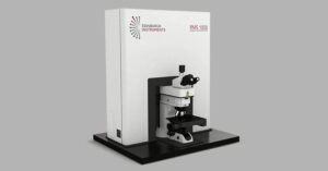
The various painted regions on the badge contain different chemical compounds that give the pigments their colour, and Raman mapping is a great technique for analysing each pigment and showing their distribution across the badge’s surface. To highlight the locations of the various pigments across the logo, and to represent the complex 3D topography of the badge, we analysed the sample with an RMS1000 Confocal Raman Microscope equipped with a 785 nm laser in Surface Mapping mode, Figure 1B. The result is a 3D surface image of the badge, showing the raised nature of the logo relative to the background and the Raman response of the differently painted regions. The corresponding spectra in Figure 1C show each region’s unique Raman fingerprint and the peaks highlighted were the ones used to build the image.
 Figure 1. Surface Mapping of Edinburgh Rugby 150th Anniversary pin badge using the Edinburgh Instruments RMS1000 Confocal Raman Microscope.


