Welcome to Edinburgh Instruments newest blog celebrating our work in Raman, Photoluminescence, and Fluorescence Lifetime Imaging. Every month we will highlight our pick for Map of the Month to show how our spectrometers can be used to reveal all the hidden secrets in your samples.
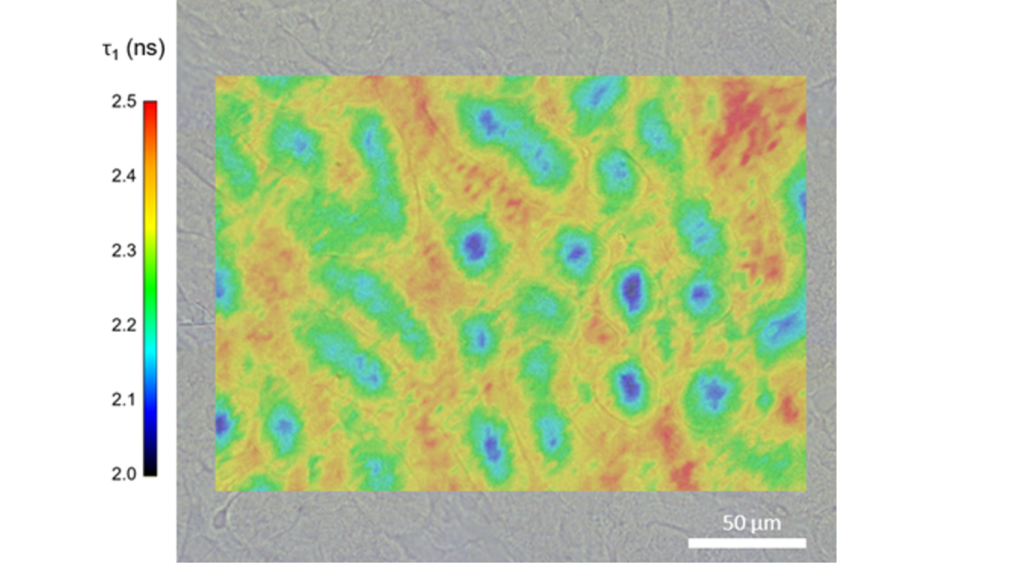
Studying animal tissue is an important tool for scientists to learn more about the effects of treatments for illnesses in humans. Mice have long served as the species of interest for biomedical research thanks to their genetic, physiological, and anatomical similarity to humans. Mice kidney samples are used to study renal biology covering topics such as acute kidney injury to chronic kidney disease, both of which are extremely useful for better understanding renal failure in humans. Fluorescence lifetime measurements are sensitive to tissue microenvironment, it’s a robust technique which, when coupled to a microscope, can provide high quality imaging on tissue samples. Here we demonstrate florescence lifetime imaging (FLIM) on a sample of mouse kidney, taken with our new MicroPL Upgrade. The FLIM map was acquired on a FLS1000 Photoluminescence Spectrometer with an EPL-485 pulsed diode as the excitation source and a high-speed PMT as the detector. The area was mapped to measure the lifetime of the 488 WGA dye in the sample, this dye stains the glycoproteins present in the cell membrane. Areas which have shorter lifetimes (blue) are the cell nuclei as the dye is not present here, the areas with longer lifetimes (red & orange) have been stained by the dye. These structures are called glomeruli which are responsible for filtering waste products from the blood.

Find out more about the FLS1000 and Micro-PL Upgrade. If you would like to talk to one of our sales team and find out how the FLS1000 and Micro-PL Upgrade can be used within your own research, please contact us.


