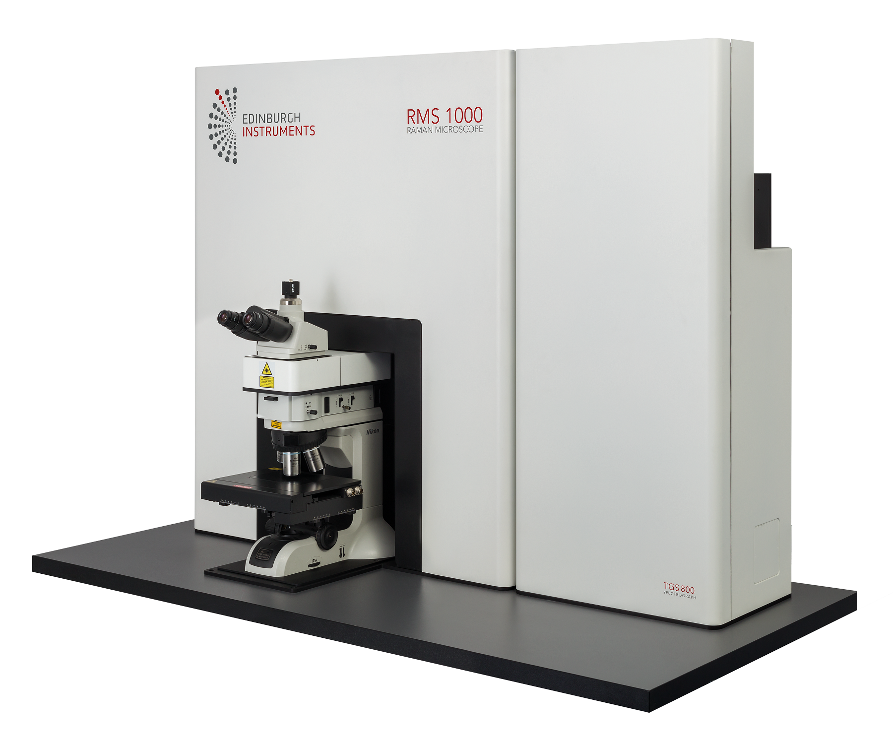
Skip to content
  • News
  • Events
  • eBooks
  • Blog
  • Careers
  • Contact
  • News
  • Events
  • eBooks
  • Blog
  • Careers
  • Contact
KNOWLEDGEBASE
  • About Us
  • Products

    Fluorescence Spectrometers

    • FLS1000 Photoluminescence Spectrometer
    • FS5 Spectrofluorometer
    • LifeSpec II Lifetime Spectrometer
    • Mini-tau Lifetime Spectrometer

    Raman Microscopes

    • RM5 Raman Microscope
    • RMS1000 Multimodal Confocal Microscope

    UV-Vis Spectrophotometers

    • DS5 Dual Beam

    Transient Absorption

    • LP980 Transient Absorption Spectrometer

    FTIR Spectrometers

    • IR5 FTIR Spectrometer

    Lasers and LEDs

    • Pulsed Lasers
    • Gas Lasers
    • Customisation Options
    View All Products
  • Techniques
  • Applications
KNOWLEDGEBASE
Edit Content
  • About Us
  • Products
  • Techniques
  • Applications
  • Knowledgebase
  • eBooks
  • News
  • Events
  • Blog
  • Careers
  • Contact Us

RMS1000

Multimodal Confocal Microscope

Elevate your research beyond Raman with the RMS1000: Its open architecture design allows for extensive customisation and upgrades, covering every application need, no matter how complex.

RMS1000

Multimodal Confocal Microscope

  • Raman and Beyond: Multimodal Raman microscope for imaging techniques such as FLIM, PLIM, SHG, and more, to take your research to the next level
  • Laser Flexibility: Up to five internal computer-controlled lasers for standard Raman and photoluminescence spectral measurements and external laser ports for advanced techniques e.g. UV, pulsed lasers, femtosecond
  • Dual Spectrograph: Upgradable to have a second, long focal length spectrograph, for unparalleled resolution
  • Multi-Detector Capability: up to four detectors including CCDs, EMCCDs, InGaAs, PMTs, for comprehensive spectral analysis.
  • Ramacle®: Unifying Raman and multimodal experiments with modern, intuitive software
View demonstration video

Send us your samples, and we can measure them live in an online demonstration

Contact us to arrange an online demonstration

Laser Flexibility

Up to five internal lasers and additional external laser ports for a range of lasers

Multi-detector

Up to four detectors including CCDs, EMCCDs, InGaAs, and PMTs for all techniques and applications

Dual Spectrograph

Standard compact and long focal length spectrographs available for ultimate resolution, sensitivity, and stray light rejection

Raman and Beyond

Multimodal confocal microscope with techniques beyond Raman, such as FLIM, PLIM, and SHG

Future Proof

Designed to grow, upgrade, and evolve with your research needs

Truly Confocal

Multi-position pinhole and slit for high spatial resolution, fluorescence and background rejection, and application optimisation
Click Here to complete an enquiry form for this product...

"*" indicates required fields

Country*
Do you have specific requirements for your instrument?*

Optional Information

What are your sample type(s) and material(s)?
Do you require mapping?
Do you require polarisation/anisotropy measurements?
Are you interested in fluorescence lifetime measurements?
Do you require temperature control?
Would you like to request a demo?

Consent

Consent*
Newsletter Subscription
Edinburgh Instruments will send you updates featuring application & technical notes, product news, exhibition announcements and other marketing activities.
This field is for validation purposes and should be left unchanged.

PRODUCT DESCRIPTION

Edit Content

The RMS1000 is a Multimodal Confocal Raman Microscope built for adaptability and designed to grow, upgrade, and evolve with your research needs.

Built without compromise, this premium research tool offers unmatched specifications and ease of use. Its unique design allows full optimisation to any Raman microscopy application. With versatile laser, detector, and spectrograph options, the RMS1000 is perfect for any laboratory, finding applications in bioscience, nanomaterials, pharmaceuticals, semiconductors, geology, and more.

The RMS1000 is more than a Raman-only system. Harnessing Edinburgh Instruments’ extensive expertise in photoluminescence, we’ve integrated advanced fluorescence techniques to create a user-friendly, multimodal instrument. Seamlessly operate Raman and fluorescence lifetime imaging (FLIM) in a single software package, making RaFLIM® data acquisition from the same point both easy and intuitive.

Expanding on Raman and FLIM, the RMS1000 is exceptionally versatile, expandable to integrate a multitude of advanced techniques. Unlock precise imaging across various techniques with our multimodal system, featuring Raman, photoluminescence (PL), fluorescence life time imaging (FLIM), phosphorescence lifetime imaging (PLIM), multiphoton techniques, and antibunching capabilities.

The RMS1000 is a cutting-edge Multimodal Confocal Raman Microscope renowned for its adaptability and ease of upgrade. Its open architecture design facilitates seamless upgrades without compromising the high specifications of the core instrument. Built to adapt to your research needs, it integrates advanced techniques ensuring precise and comprehensive imaging capabilities across various scientific disciplines.

PRODUCT VIDEO

PRODUCT DETAILS

SOFTWARE

Ramacle® is an all-encompassing software package written for complete instrument control and data handling for all multimodal techniques on the RMS1000.

Just like the hardware, the software operating the RMS1000 adapts to your research need. Ramacle controls all RMS1000 functions with a straightforward design concept. It focuses on all modern Raman spectroscopy applications, and beyond. For example, running Raman and FLIM maps easily in one comprehensive platform.

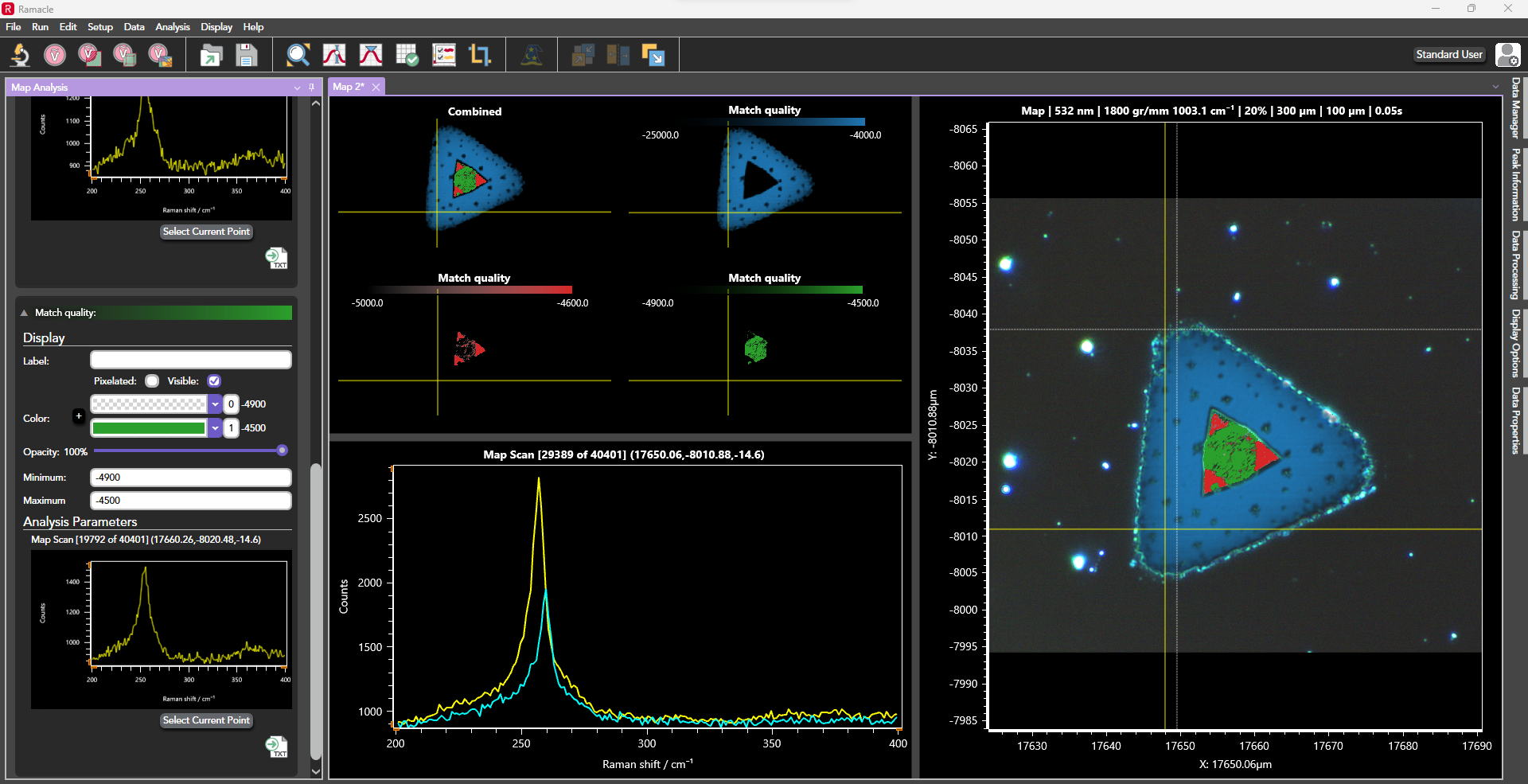
Figure 1: Ramacle software interface

With Ramacle, you can easily navigates from microscope set up to measurement set up and offers computer-controlled measurement conditions such as laser excitation, grating, pinhole size etc. On the RMS1000, Ramacle comes with standard data acquisition methods such as single measurements, multiple and accumulated scans, kinetic scan, as well as a comprehensive suite of mapping techniques e.g. 2D, 3D, SurfMAP, and FastMAP. Upgrades and accessories such as temperature stages and multiwell plates unlock additional software features.

MEASUREMENT EXAMPLES

Raman Mapping
SurfMAP®
RaFLIM®
Multimodal Techniques  
Raman Mapping

2D mapping and 3D mapping allow the user to see distribution of components, as well as revealing areas under high stress or strain, and defects. If using short exposure times FastMAP® is also available to reduce total acquisition times. ​

2D Raman mapping showing nanocrystalline silicon and strain (pink, yellow, red) in a defective wafer

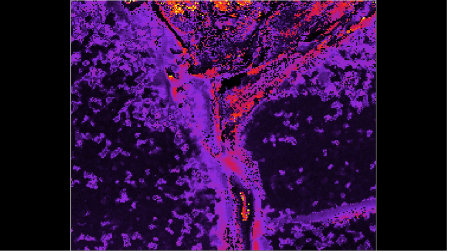
Figure 1: 2D Raman mapping showing nanocrystalline silicon and strain (pink, yellow, red) in a defective wafer

3D Raman map of a pharmaceutical oil-in-water emulsion with TiO2 inclusions

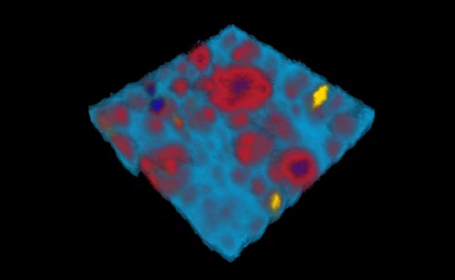
Figure 2: 3D Raman map of a pharmaceutical oil-in-water emulsion with TiO2 inclusions

SurfMAP®

SurfMAP® is a tool which allows accurate mapping of uneven surfaces giving the user assurance that their measurements are in focus and data is reliable. Ramacle builds a surface image which can be viewed in 2D and in 3D topography view. The software then acquires the spectral map moving the stage in X, Y, and Z for perfect laser focus.

Raman SurfMAP® of carbon fibres tracking the D and G bands

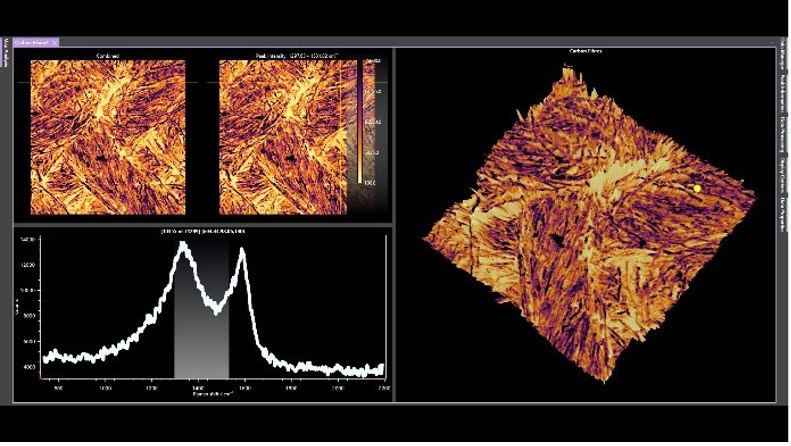
Figure 1: Raman SurfMAP® of carbon fibres tracking the D and G bands

Raman SurfMAP® of antipsychotic drug tracking the D and G bands

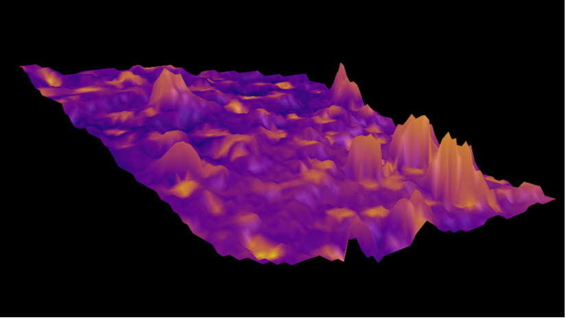
Figure 2: Raman SurfMap of an antipsychotic pharmaceutical drug

RaFLIM®

RaFLIM is a standout technique on the RMS1000. Combining Raman and FLIM to create maps of the same area, with the same spatial resolution (laser dependent). Operating measurement acquisition and data analysis in Ramacle for ease of use and continuity.

average lifetime and fluorescence intensity image of a Convallaria Rhizome section stained with Acridine Orange. FLIM reveals variation in lifetime across lignified and pectin rich cell walls (2 exponential tail fitting)

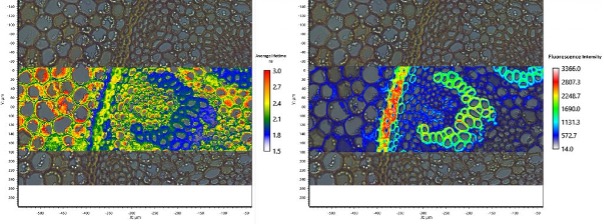
Figure 1: Average lifetime and fluorescence intensity image of a Convallaria Rhizome section stained with Acridine Orange. FLIM reveals variation in lifetime across lignified and pectin rich cell walls (2 exponential tail fitting).

Biochemical analysis of woody plant cell walls using our multi-modal micro-spectroscopy approach

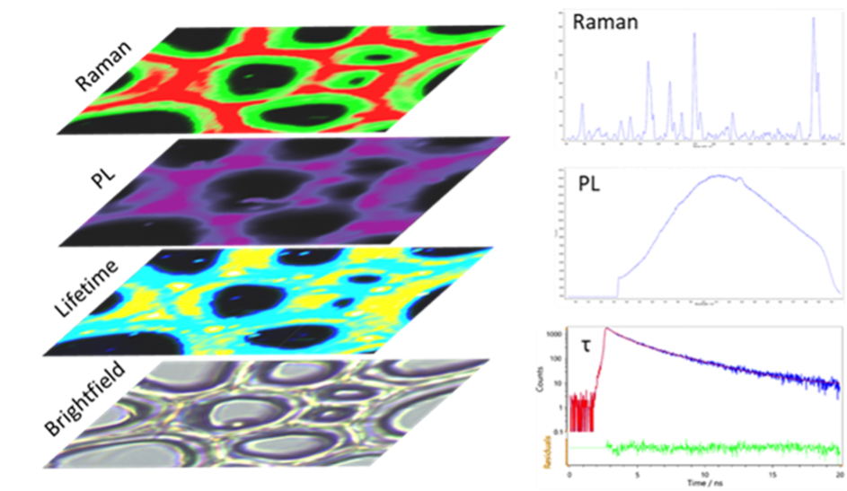
Figure 2: Biochemical analysis of woody plant cell walls using our multi-modal micro-spectroscopy approach.

Multimodal Techniques  

The RMS1000’s external laser port unlocks limitless potential for advanced techniques like multiphoton imaging. Techniques such as two-photon excited fluorescence (2PEF) and second harmonic generation (SHG) are ideal for studying biological samples. Both 2PEF and SHG demand extremely high excitation intensity, provided by a mode-locked femtosecond pulsed laser.

Mouse intestine stained with Alexa Fluor® 568

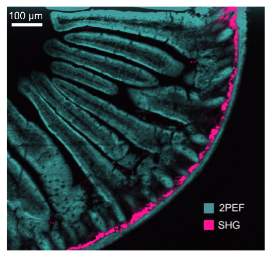
Figure 1: Mouse intestine stained with Alexa Fluor® 568

Photon lifetime imaging of mouse kidney sample

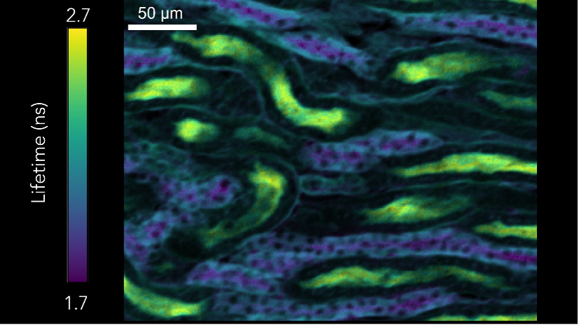
Figure 2: Photon lifetime imaging of mouse kidney sample

The sample of the mouse intestine (Figure 1) stained with Alexa Fluor® 568. Simultaneous measuring of spectral 2PEF and SHG from Alexa Fluor® 568 dye and fibrillar collagen respectively. Measurement using femtosecond laser and CCD camera. Using the same laser, two photon lifetime imaging was carried out an a sample of mouse kidney (Figure 2). This time a Hybrid Photodetector was used with TCSPC.

2D materials are also often studied with different measurement techniques, the RMS1000 can easily be configured to carry out all imaging techniques on the same sample area providing a full suite of complimentary information. Raman and PL images were collected using a 532 nm laser, whilst the SHG was acquired using a femtosecond laser.

WSe2 crystal. The darkfield (DF) image reveals areas of different heights. Raman, PL, and SHG images can be used correlatively to identify layer number, and highlight areas of stress, strain, dopants, and defects.

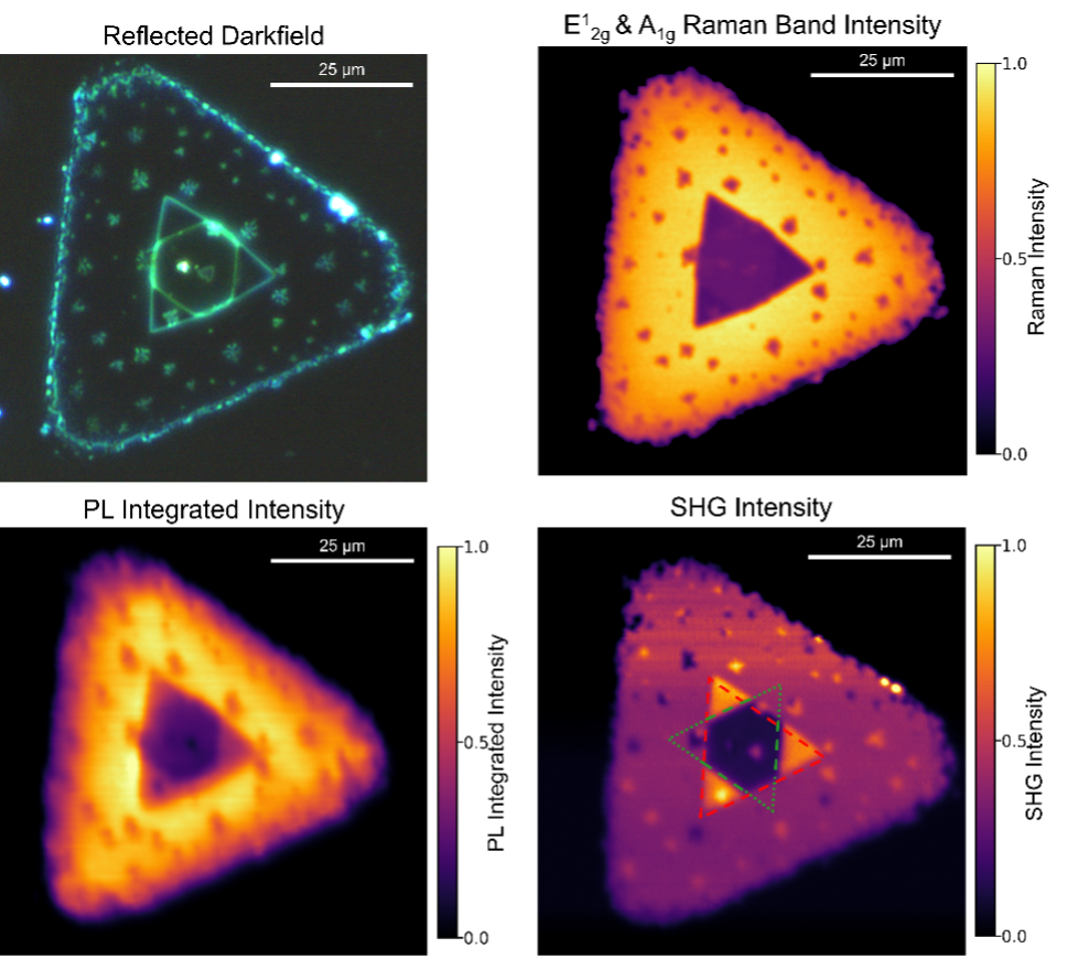
Figure 3: Sample of WSe2 crystal. The darkfield (DF) image reveals areas of different heights. Raman, PL, and SHG images can be used correlatively to identify layer number, and highlight areas of stress, strain, dopants, and defects

Customer Testimonials

“The optomechanical design of the RMS1000 is open and intuitive, enabling us to easily adjust and customize it to meet our different Research groups needs efficiently”
RMS1000 Testimonial
Dr Andrés De Luna Bugallo, UNAM, Mexico

Options and Upgrades

Lasers
SpectrographsRaman Microscope Laser Upgrade
Detectors
Microscope Accessories
Sample Stages
Accessories and Laser Safety
Lasers

The RMS1000 is built with flexibility in mind. A choice of excitation lasers (internal or external to the system) and associated laser rejection filters (both edge and notch) are available. Additionally pulsed diode lasers such as our EPL series can be added if fluorescence lifetime measurements are required.

Raman Microscope Laser Upgrade

SpectrographsRaman Microscope Laser Upgrade

The RMS1000 can be fitted with both standard and long focal length spectrographs. This allows the system to be configured for ultimate spectral resolution, maximum optical throughput, low wavelength and ultraviolet spectral ranges, or for highly sensitive measurements.

Raman Microscope upgrade - Spectrograph

Detectors

A choice of CCD, EMCCD and InGaAs detectors are available with a maximum of four detectors being integrated per system. For fluorescence lifetime measurements, single photon counting detectors can also be integrated.

RM5 Raman Microscope Detector

Microscope Accessories

The RMS1000 uses either an upright or inverted microscope platform which is compatible with all standard microscopy applications.

Brightfield, darkfield, polarised light, differential interference contrast (DIC) and fluorescence accessories are all available. A large choice of high-quality microscope objectives, high-performance visualisation cameras, collimators and illuminators can be added to the microscope at any time.

Raman Microscope Accessories for upgrade

Sample Stages

Manual or motorised stages are available. The motorised stage allows automated XYZ Raman and fluorescence maps to be obtained and generated through Ramacle. Additional sample stages are compatible with theRMS1000, such as temperature stages, electrochemical cells, and diamond anvil cells.

Raman microscope sample holder - upgrade

Accessories and Laser Safety

Other accessories such as a polarisation kit, cuvette holder, and a Class I laser safety enclosure are also available to further expand the capabilities, flexibility and safety of your RMS1000 system. Coupling to other fluorescence spectrometers is also possible.

Raman Microscope laser safety upgrade

TECHNICAL SPECIFICATIONS

RMS1000
RMS1000
Specifications
LASERSUp to 5 integrated narrow-band lasers: 532 nm, 638 nm, 785 nm typically used
Additional lasers from UV to NIR are available. External lasers can be integrated
Laser selection is fully computer-controlled
Associated laser rejection filters included, fully computer-controlled
SPECTROGRAPHSWavelength Range200 nm - 2,200 nm
Gratings5-position grating turrets
SlitsContinuously adjustable, fully computer-controlled
SPECTRAL RESOLUTIONFrom <0.1 cm-1 (depending on grating, laser and CCD selection)
SPATIAL RESOLUTIONXY (lateral), Z (axial)0.25 µm, <1 µm (depending on laser and microscope objective)
SPECTRAL RANGE5 cm-1 * - 30,000 cm-1 (* with low wavenumber attachment)
CONFOCAL IMAGINGAdjustable confocal pinhole, fully computer-controlled
DETECTORSCCD DetectorHigh sensitivity ultra low noise spectroscopy CCDs
1650 x 200 pixels, TE-cooled -60°C (standard)
2000 x 256 pixels, TE-cooled -60°C (enhanced sensitivity / spectral range)
Optional Second DetectorEMCCD detector, 1600 x 200 pixels, TE-cooled -100°C (fast response time)
InGaAs array, 1024 pixel, TE-cooled -90°C, up to 2,200 nm
SOFTWARERamacle®Comprehensive all-in-one, intuitive software package
OptionalChemometric, spectral library packages
FLUORESCENCESpectralWith low resolution grating and integrated CCD
LifetimeWith picosecond pulsed lasers, TCSPC electronics, fast photon counting detectors
Spectral and lifetime fluorescence mapping is also available
LASER SAFETYWithout Laser EnclosureClass 3B (depending on external laser source)
With Laser EnclosureClass 1
DIMENSIONSW x D x HFrom 975 mm x 610 mm x 1170 mm
WeightFrom 114 kg

PRODUCT BROCHURE

APPLICATION NOTES

  • Multimodal Raman, Photoluminescence & SHG Imaging of CVD Grown WSe2
  • 3D Raman Mapping of a Transdermal Patch
  • Raman and Photoluminescence Characterisation of GaN
All Application Notes

DOWNLOADS

  • RMS1000 Raman Microscope Brochure
Enquire Now

Keep up to date with the latest from Edinburgh Instruments

Join our mailing list and keep up with our latest videos, app notes and more!

LOCATION:
  • Edinburgh Instruments Ltd.
    2 Bain Square, Kirkton Campus, Livingston, EH54 7DQ.
  • sales@edinst.com
  • +44 1506 425 300
ABOUT:
  • About Us
  • Techniques
  • Applications
  • Knowledgebase
  • About Us
  • Techniques
  • Applications
  • Knowledgebase
PRODUCTS:
  • Fluorescence Spectrometers
  • Raman Microscopes
  • UV-Vis Spectrophotometers
  • Transient Absorption
  • FTIR Spectrometers
  • Lasers and LEDs
  • Customisation Options
  • Software
  • Upgrades
  • All Products
  • Fluorescence Spectrometers
  • Raman Microscopes
  • UV-Vis Spectrophotometers
  • Transient Absorption
  • FTIR Spectrometers
  • Lasers and LEDs
  • Customisation Options
  • Software
  • Upgrades
  • All Products
LEGALS:
  • News
  • Events
  • Blog
  • Careers
  • Contact Us
  • Terms and Conditions
  • Privacy Policy
  • News
  • Events
  • Blog
  • Careers
  • Contact Us
  • Terms and Conditions
  • Privacy Policy
SOCIALS:
Youtube Linkedin X-twitter Facebook
©2024 Edinburgh Instruments. Registered in England and Wales No: 962331. VAT No: GB 271 7379 37
Manage Consent
To provide the best experiences, we use technologies like cookies to store and/or access device information. Consenting to these technologies will allow us to process data such as browsing behaviour or unique IDs on this site. Not consenting or withdrawing consent, may adversely affect certain features and functions.
Functional Always active
The technical storage or access is strictly necessary for the legitimate purpose of enabling the use of a specific service explicitly requested by the subscriber or user, or for the sole purpose of carrying out the transmission of a communication over an electronic communications network.
Preferences
The technical storage or access is necessary for the legitimate purpose of storing preferences that are not requested by the subscriber or user.
Statistics
The technical storage or access that is used exclusively for statistical purposes. The technical storage or access that is used exclusively for anonymous statistical purposes. Without a subpoena, voluntary compliance on the part of your Internet Service Provider, or additional records from a third party, information stored or retrieved for this purpose alone cannot usually be used to identify you.
Marketing
The technical storage or access is required to create user profiles to send advertising, or to track the user on a website or across several websites for similar marketing purposes.
Manage options Manage services Manage {vendor_count} vendors Read more about these purposes
View preferences
{title} {title} {title}