Raman microscopy is a powerful technique for identifying the source of forensic samples, such as paint traces. As a non-destructive method of forensic analysis, evidence samples are not compromised during investigation. During forensic investigations, evidence samples are often compared to a database of known specimens for identification. In this Application Note, we show how the RM5 can be used to differentiate between different samples and how spectral library searching allows for the matching of paint samples within seconds.
Forensics teams play a crucial role in crime investigations, routinely analysing trace evidence to implicate suspects or determine commonalities between different crimes. Among the most easily identifiable pieces of trace evidence are pigments and dyes from chips of paint, synthetic fibres, or inks.1 These sample types are used in comparative forensic analysis, whereby a ‘questioned’ specimen is submitted to a forensic laboratory and compared with known specimens to identify common characteristics. Investigators could then use these analyses to build patterns between events, objects or individuals. For example, comparative analysis of paint traces could be used to identify the transfer of pigments between vehicles during a collision, objects such as crowbars or screwdrivers used to break into windows, cars, or safes, or the source of spray paint used in graffiti. This can allow examiners to track vehicle types in hit-and-run accidents, catch thieves who unknowingly transfer paint from the robbery site onto any implements they use and identify the seller of spray paints used in cases of vandalism or hate speech, Figure 1.
Comparative analysis of these samples must be performed with extremely high chemical specificity to discriminate between different pigments of similar hues. It must also be capable of non-destructively analysing microscopic traces so that they can later be analysed using corroborative techniques. Raman microscopy meets both criteria because of its high spectral resolution, the available sub-micron range spatial resolution, and the fact that no sample preparation is required. Importantly, it is non-destructive, ensuring the preservation of evidence. It excels at analysing very low concentrations of paint because of the high molecular symmetry and resonance enhancements of the pigment molecules, which means that these samples often produce intense scattering, as is shown in Figure 1.2 It is also highly versatile and can be applied to detecting other common types of evidence of interest to forensic laboratories, including drugs and explosives. This Application Note shows how the Edinburgh Instruments RM5 Confocal Raman Microscope can be used to analyse paint traces in a forensic laboratory setting.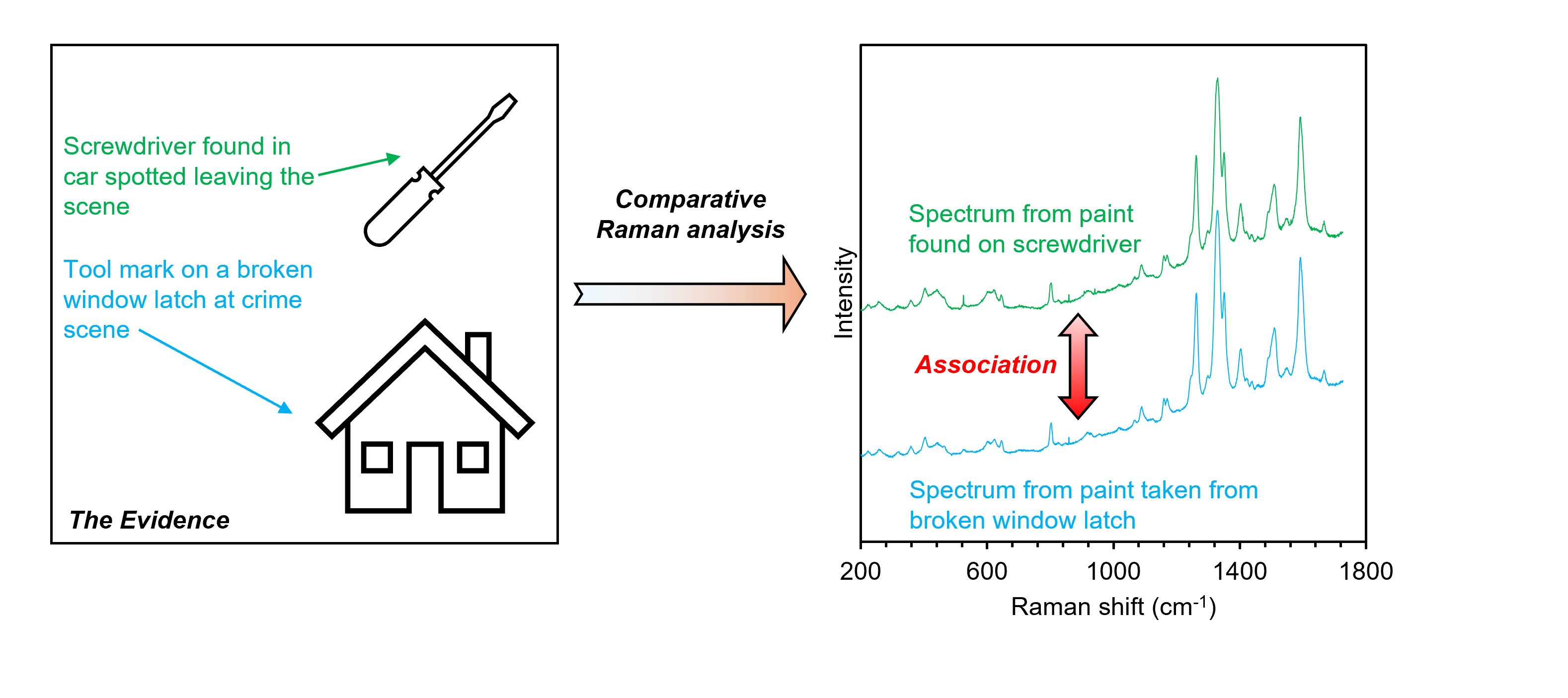 Figure 1. Example of where comparative Raman analysis of paint traces could be used for forensic crime scene investigations.
Raman spectroscopic measurements were performed on an RM5 Raman Microscope equipped with 532 nm, 638 nm, and 785 nm lasers and a CCD detector, Figure 2. The RM5’s Ramacle® software fully automates laser control, meaning that measurement parameters can be changed without disturbing the sample between measurements. The software-controlled laser attenuator within the RM5 was also used to keep the power reaching the sample low and prevent damage.
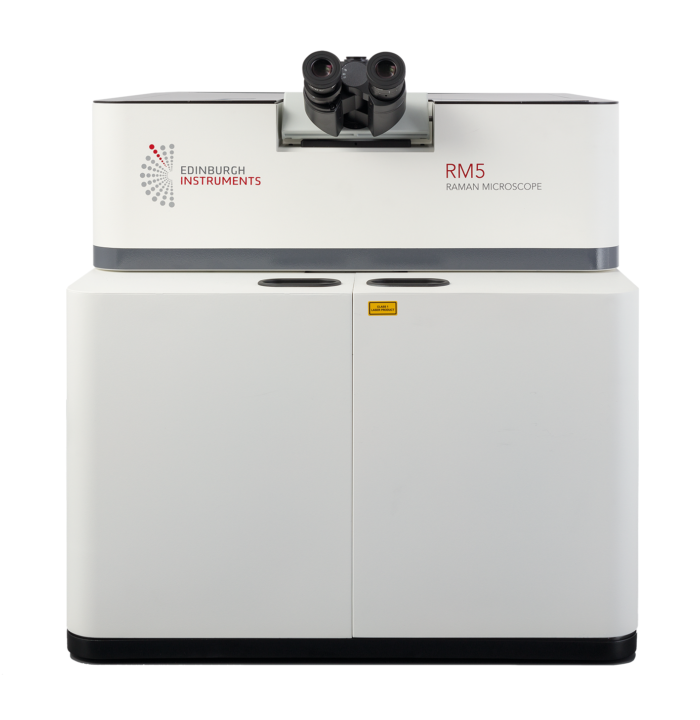 Figure 2. Edinburgh Instruments RM5 Raman Microscope.
Paint generally contains pigments, binders, solvents, and other additives. Pigments can either consist of brightly coloured natural inorganic compounds, ubiquitous in historical art pieces as shown in our artwork and archaeology Application Note, or synthetic organic compounds, which are more commonly used now. However, inorganic titanium dioxide or carbon-based pigments are still commonly found in white and black paints, respectively. The organic compounds used in paints generally contain highly polarisable conjugated carbon chains or aromatic functional groups, which produce distinct and intense Raman signatures. Therefore, the technique is highly effective at detecting and differentiating between different paint compounds. To demonstrate this, various paint samples were analysed using the RM5.
The first samples analysed were two paint traces that looked similar under widefield microscopy, suggesting a common origin, Figure 3a. Raman spectra were recorded from multiple points on both traces, Figure 3b. While both traces share some Raman signatures (orange peaks), there are significant differences. Trace 1 has several unique signatures (green peaks), and the intensity of certain bands differs between the traces. For example, the band at 1597 cm-1 is more intense in Trace 1, while the band at 1316 cm-1 is stronger in Trace 2. This indicates that the traces contain common compounds in different amounts, with Trace 1 having additional compounds. This study shows Raman spectroscopy is effective for distinguishing visually similar samples.
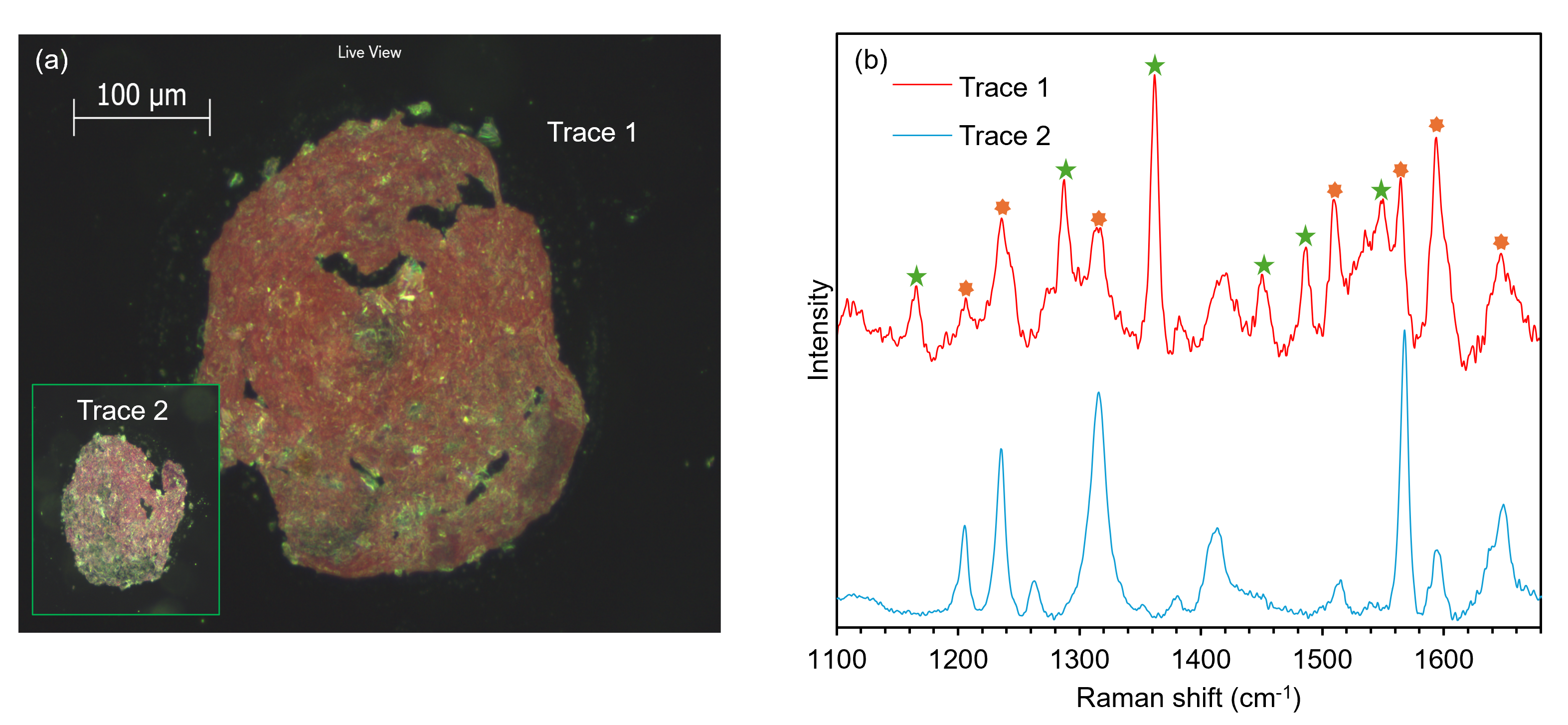
Figure 3. Comparative Raman spectroscopic analysis of paint traces.
Next, a yellow spray paint was analysed using different wavelength lasers for excitation. Raman spectra obtained using different lasers are usually qualitatively similar, but the intensities of the bands will differ when recorded over the same acquisition times because of the negative . For this sample, however, there were notable qualitative differences in the raw spectra recorded at 532 nm and 785 nm, Figure 4. The Raman spectrum recorded at 532 nm is dominated by intense bands at 1262 cm-1, 1329 cm-1, and 1592 cm-1. These bands are still present and intense in the spectrum recorded at 785 nm, but two broad Raman bands at 450 cm-1 and 620 cm-1 were also detected, which were assigned as the rutile form of titanium dioxide.3 Note that the 785 nm spectrum was recorded over a longer acquisition time than the 532 nm spectrum. These differences were attributed to the resonance enhancement of the yellow pigment at 532 nm, which meant the dominant spectral signatures of the chromophore obscured the rutile.
In the forensic analysis of pigments, resonance enhancements must be considered in order to avoid erroneous mismatches based on qualitative differences in spectra recorded under different measurement conditions. For example, if the excitation wavelengths were not entered into the metadata of a Raman spectral library, it could be concluded that the spectra in Figure 4 belonged to different paint samples because of the varying contributions of the organic pigment and the rutile.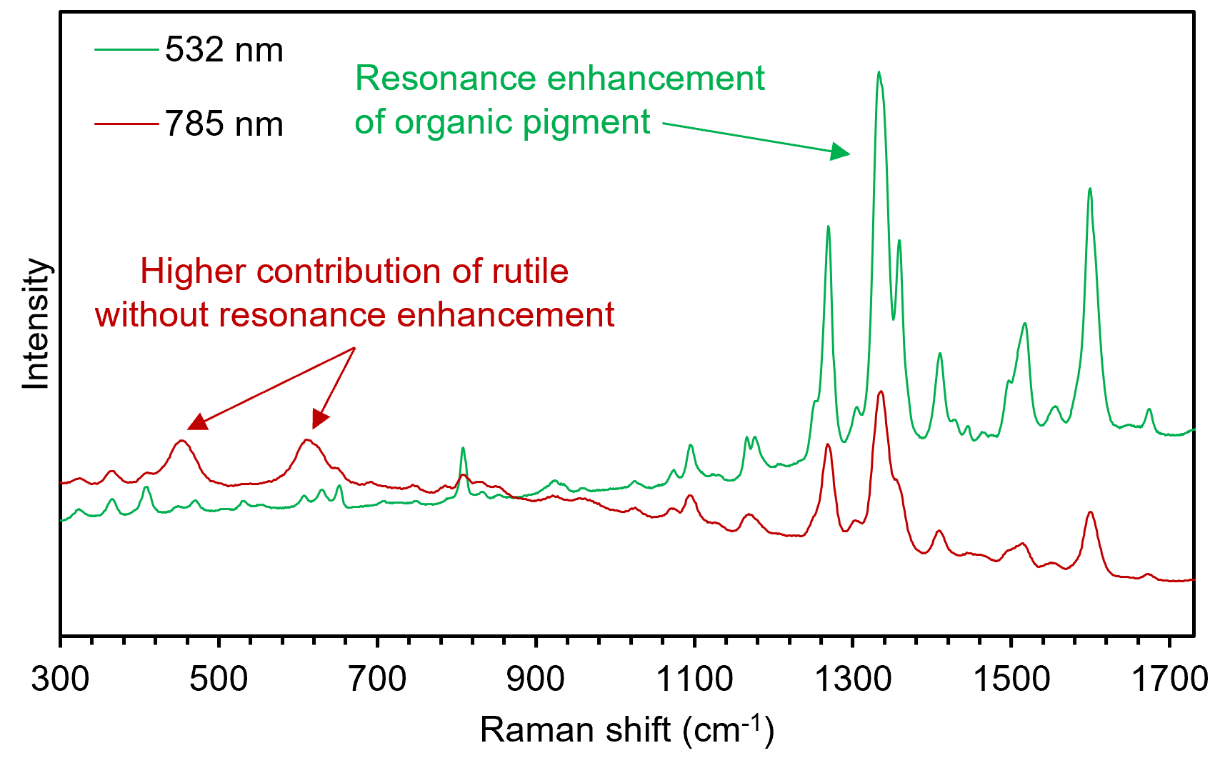 Figure 4. Multiple wavelength Raman spectroscopic analysis of yellow spray paint.
In forensic Raman spectroscopy, recorded spectra are often compared against a spectral library to find matches that can provide case-relevant connections. Ramacle allows seamless transfer of a spectrum into KnowItAll, which offers numerous spectral libraries and options for user-built libraries. By clicking the KnowItAll icon in Ramacle, the spectrum is analysed through the database in seconds.
A sample of black spray paint was analysed and identified, Figure 5. Using the intuitive export function from Ramacle to KnowItAll the spectrum was rapidly identified as carbon black, with a 95% match. This information is then returned and stored in Ramacle with all other metadata. The dominant features in the spectrum were large bands at 1338 cm-1 and 1587 cm-1, characteristic of the D and G bands of amorphous carbon. It has been demonstrated in the literature that different carbon-based black pigments vary subtly in the position, width, and relative intensity of the D and G bands. Analysing various black paint brands and creating a spectral library from these samples could help identify where a black spray paint used by a perpetrator was sold.4
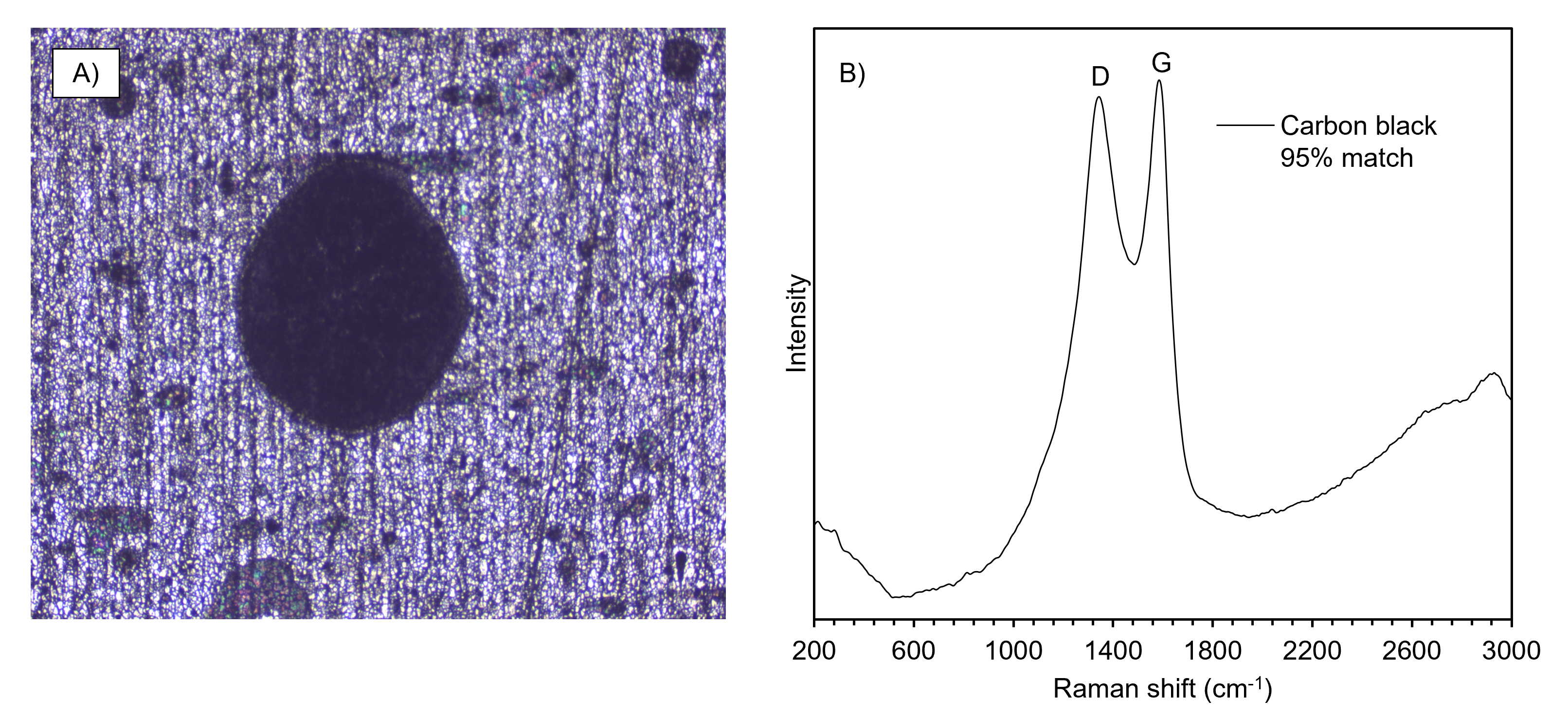
Figure 5. Raman spectral analysis and KnowItAll identification of carbon black spray paint.
This Application Note shows how the Edinburgh Instruments RM5 Confocal Raman Microscope can be used to analyse paint traces in a forensic laboratory setting. Raman microscopy is a powerful technique for analysing and identifying pigments in paint traces.
