Under extreme conditions, perovskites can behave quite differently. By studying perovskites under these conditions can unlock novel properties and demonstrate new applications. Here, we investigate spectral and time-resolved photoluminescence of halide perovskites under high pressure using the RMS1000 and a diamond anvil cell.
Halide perovskites are a promising class of semiconductors for optoelectronic applications. Research on halide perovskite has focused on development methods, composition engineering, and post-treatment techniques, with the aim of improving the quality and enhancing the properties of the materials.1 Significant progress has been made; halide perovskites are now being produced with ideal bandgaps of 1.1 to 1.5 eV for photovoltaic applications and very low numbers of crystalline defects, which has resulted in solar cells that can operate over long operating times with high power conversion efficiencies.2,3 Innovation is still required in order for these materials to be further improved and used more widely.
One technique for obtaining new material information and revealing novel properties is high-pressure photoluminescence spectroscopy. Under high pressure, the distances between atoms within a crystalline lattice decrease, altering their interactions and properties. Shortening the distance between atoms increases orbital overlap and influences the bandgap of the material. Further, new phases can form in the crystalline lattice, which could also exhibit unique properties.
In this Application Note, we show how the Edinburgh Instruments RMS1000 Confocal Microscope with the fluorescence lifetime imaging (FLIM) upgrade can be used to perform spectral and time-resolved photoluminescence (PL) analysis of halide perovskite crystals under high pressure in a diamond anvil cell (DAC).
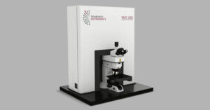
(PEA)2PbBr4 crystals in a DAC were analysed using an RMS1000 Confocal Multimodal Microscope equipped with a 405 nm HPL picosecond pulsed diode laser, a back-illuminated CCD detector for spectral measurements, a high-speed hybrid photodetector (HSHPD) for time-resolved measurements, and TCC2 photon counting electronics for time-correlated single photon counting (TCPSC), Figure 1. The diamond anvil cell was placed on the motorised stage of the RMS1000 and brought into focus under a 10X 0.3 NA objective. To acquire PL spectra of the crystals, the HPL was used as a quasi-CW source by selecting the highest repetition rate, and emission was detected on the CCD detector after dispersion by a 300 gr/mm grating. For lifetime measurements, the HPL was used to excite the sample with a repetition rate of 1 μs, the PL was detected using the HS-HPD, and decays were recorded using TCSPC.
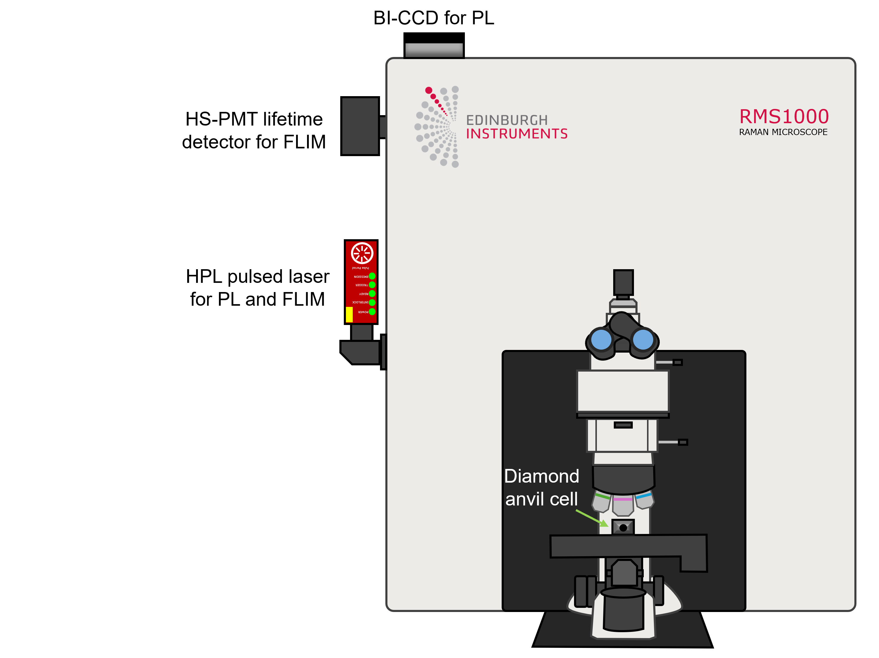
Figure 1. Edinburgh Instruments RMS1000 Multimodal Microscope for high-pressure halide perovskite spectral and time-resolved photoluminescence analysis.
DACs consist of two opposing diamond anvils with the culets facing each other, a sample chamber between the two diamonds in which the sample is immersed in a pressure-transmitting medium with a ruby crystal for measuring the chamber pressure, and a gasket for securing the sample, as shown in Figure 2.4 Pressure is applied to the sample by compressing the anvils, which is done by tightening screws on the outside of the cell. The diamond is transparent to a large range of the electromagnetic spectrum, meaning it is possible to perform a range of spectroscopic measurements on the sample, including PL, Raman, and second harmonic generation (SHG). In summary, DACs are cost-efficient, compact, and an excellent tool for fundamental high-pressure spectroscopic studies of small amounts of material.
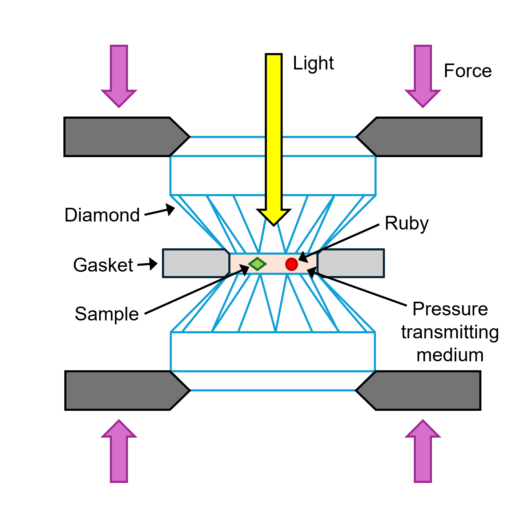
Figure 2. Diamond anvil cell for analysing crystals under high pressure.
The sample chamber in the DAC was focused under the microscope, and the halide perovskite crystals were located using brightfield microscopy, Figure 3. Figure 3a shows the sample chamber illuminated in reflection brightfield, and Figure 3b shows transmission brightfield. The transmission brightfield image provided a better optical contrast and visualisation of the perovskite crystals and the pressure calibration ruby.
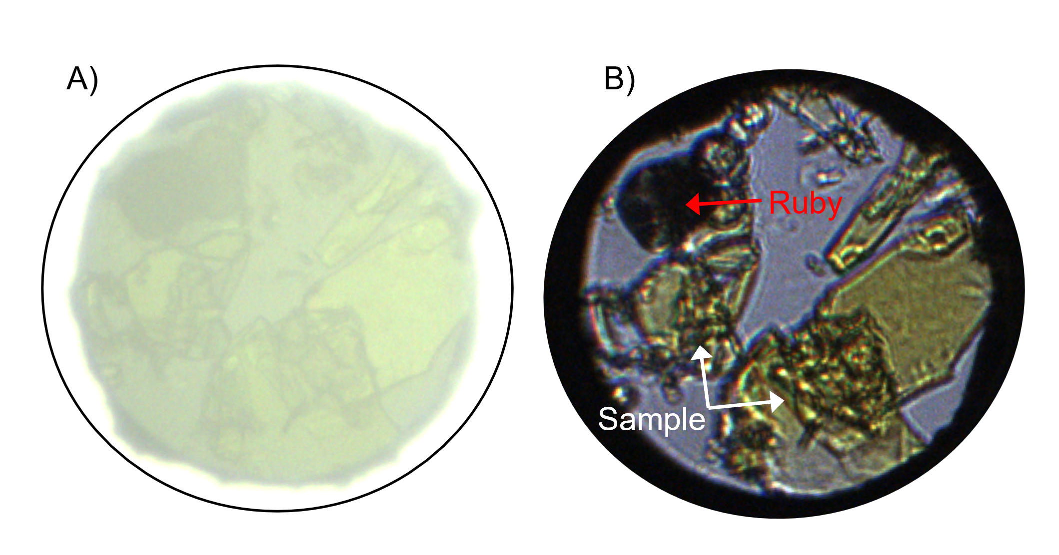
Figure 3. A) Reflected brightfield and B) transmission brightfield imaging of the DAC sample chamber.
The perovskite and ruby were then analysed using spectral PL, Figure 4. Figure 4A shows spectra recorded from the perovskite crystal and a region where no sample was present in green and blue, respectively. The acquisition times for both of these spectra was 0.01s. The perovskite crystal exhibited a broad PL emission centred at 700 nm (1.77 eV). The entire sample chamber showed a peak at 460 nm, which was therefore assigned as PL from the pressure-transmitting medium. Figure 4B shows the PL spectrum of the ruby crystal; two peaks were detected at 697 nm and 699 nm. The position of the more intense and higher wavelength PL band is typically used to calibrate the pressure in DACs, with a redshift from 694 nm indicating pressures greater than 1 atm.5
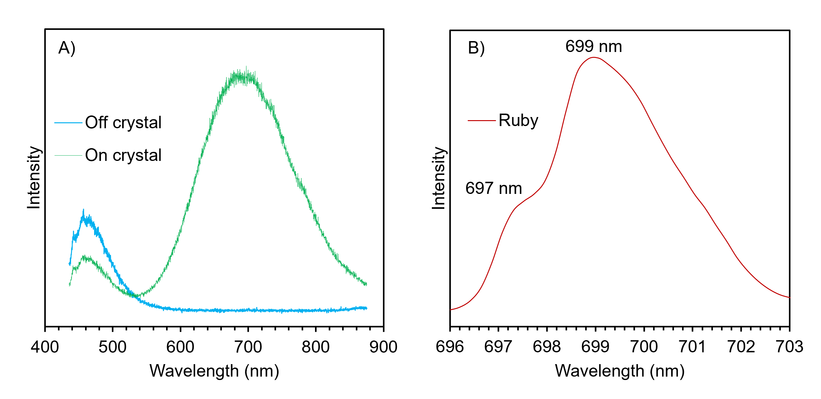
Figure 4. PL spectra of the A) perovskite and B) ruby crystals.
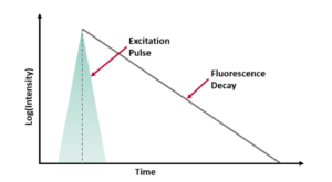
The perovskite crystals were then analysed using time-resolved PL. Lifetime decays were acquired from the perovskite crystal at a wavelength of 670 nm to avoid the ruby PL signatures using TCSPC with an EPL repetition rate of 1 μs, and the sample was mapped in X and Y. The decays at each point in the map were fit with a three-component exponential model using the Ramacle® software of the RMS1000 and the intensity-weighted average lifetime was used to create the lifetime image shown in Figure 5A. The image shows clear variation in the lifetime across the perovskite crystals. Figure 5B shows a representative decay showing the multi-exponential nature of the fit.
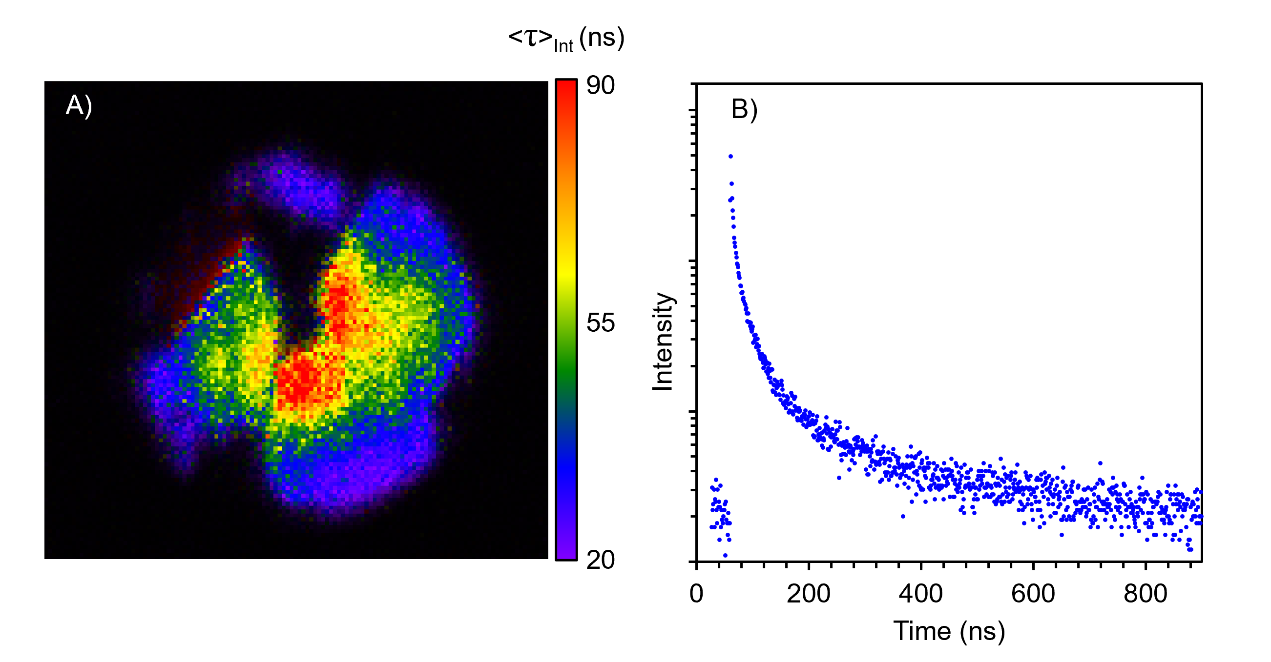 Figure 5. FLIM of the perovskite crystals.
In this Application Note, we have shown how the Edinburgh Instruments RMS1000 Confocal Multimodal Microscope with the FLIM upgrade can be used to perform spectral and time-resolved PL analysis and imaging of halide perovskite crystals subjected to high pressure in a DAC. High pressure studies of perovskite crystals are a highly promising avenue for unlocking new properties and realising novel applications. Raman and SHG analysis and imaging are also possible in DACs with the RMS1000.
References

