FLIM Microscopy involves exciting a sample with a pulsed laser and then measuring the time it takes for the fluorescence to decay after excitation. FLIM can be used in a variety of applications including:
Materials science: FLIM is used to measure the spatial variation in charge carrier lifetimes across semiconductor materials and investigate loss mechanisms.
Biological imaging: A sample is stained with one or more fluorophores where the fluorophore lifetime is influenced by various factors, such as the local environment, quenching effects, and interactions with other molecules.
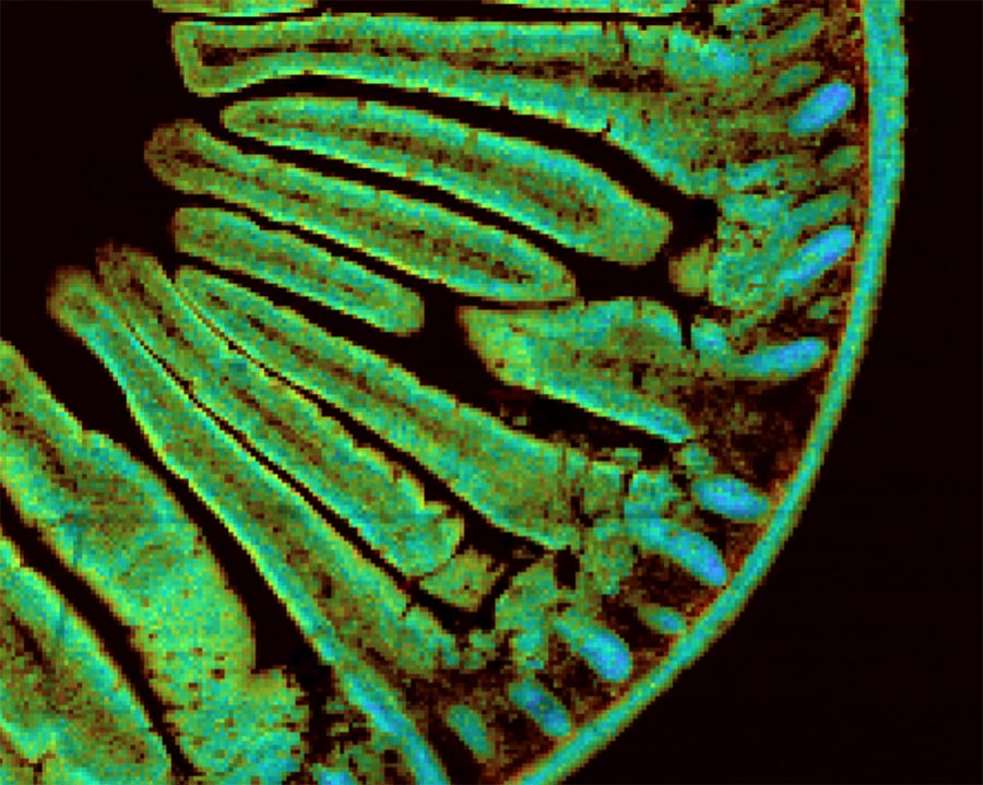
Figure 1: FLIM Imaging of mouse intestines.
FLIM Microscopy is used for lifetimes in the picoseconds to nanoseconds time range and utilises Time-Correlated Single Photon Counting (TCSPC). Phosphorescence Lifetime Imaging (PLIM) using Multichannel Scaling Photon Counting (MCS) is a complementary technique for measuring longer microsecond and millisecond lifetime images; however, both techniques are often collectively called FLIM.
Edinburgh Instruments fluorescence spectrometers and Raman Microscopes can be extended with both FLIM and PLIM capability.