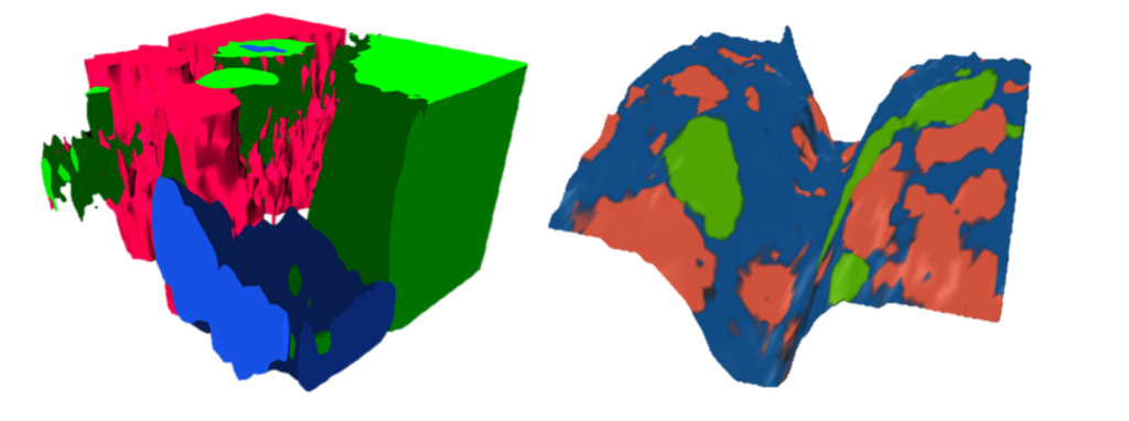
Raman microscopy merges the chemical fingerprinting power of Raman spectroscopy with high-resolution optical imaging to deliver detailed chemical visuals of materials. By detecting vibrational modes through light scattering, it offers a non-destructive method to analyse chemical composition. Learn how our RM5 and RMS1000 Raman Microscopes can enhance your research in Raman Microscopy.
In Raman microscopy, monochromatic laser light is directed onto a sample, and the scattered light is collected and analysed.
Most of the scattered light retains the same energy as the incident light (Rayleigh scattering), but a small fraction undergoes energy shifts due to interactions with molecular vibrations (Raman scattering). These energy shifts provide a molecular fingerprint that is unique to the specific bonds and structures within the sample. Detailed Raman images of the sample's chemical composition are obtained by acquiring the Raman spectra across a 2D area or 3D volume.
Raman microscopy is useful for a wide variety of sample types, including studying biological tissues, characterising semiconductor and nanomaterial layers, and analysing polymers and pharmaceuticals. Its non-invasive nature allows for rapid analysis without the need for extensive sample preparation.
